
Sub-Aortic Stenosis in Canines
- The Student Vet
- Jun 28, 2018
- 5 min read

During my work experience i encountered a patient with a grade 5 sub aortic stenosis. This inspired me to further my knowledge by researching more about this condition.
Definition and Cause:
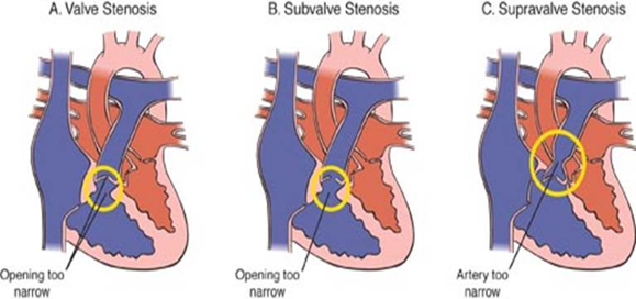
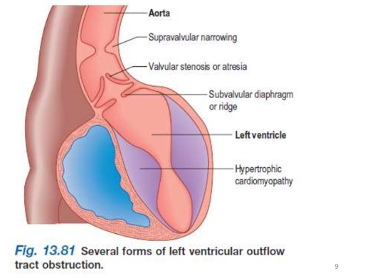
Aortic stenosis impedes normal left ventricular emptying and is caused by a narrowing at one of three locations:
1) below the aortic valve (subvalvular or subaortic)
2) at the aortic valve (valvular)
3) above the aortic valve (supravalvular).
The most common form of aortic stenosis in dogs is subaortic stenosis, caused by a ridge of fibrous tissue within the left ventricular outflow tract, just below the aortic valve. This restricts and interferes with the aortic blood flow. The aorta is the largest artery in the body and is responsible for supplying the body's cells with oxygen and glucose. It is a vital blood supply that must not be compromised. Subaortic stenosis is generally seen in large-breed dogs. Predisposed breeds include Boxers, Golden Retrievers, Rottweilers, German Shepherds, and Newfoundlands.
Subaortic stenosis is an autosomal-dominant (incomplete penetrance) congenital heart disease of dogs, characterized by a narrowing of the descending aorta below the left ventricular outflow tract of the heart.
The narrowing is due to muscular and fibrous tissue that keeps growing longer than they should. When a puppy with SAS is first born, the heart is only slightly abnormal in this area. The tissue in this subaortic area can remain only slightly abnormal, or it can continue to grow until it prevents blood from passing through the narrowed channel. The more restricted the blood flow becomes the louder the heart murmur and the more serious the problem.


Symptoms:
Usually, with SAS there will be a heart murmur but not in all cases. In mild sub-aortic stenosis, no signs are observed. In moderate and severe cases, there are symptoms such as weakness, breathing difficulty (dyspnea), fainting (syncope), and, in extreme cases, sudden death. All these symptoms are due to the strain on the heart resulting in a lack of oxygen being delivered to certain areas like the brain causing syncope. The animal, in general, will have less energy as there is decrease rate of respiration due to a compromised transportation system of glucose and oxygen.
Heart murmurs are graded based on the intensity and loudness of the murmur. The grades range from 1 to 6, 1 being barely audible with a stethoscope and 6 being clearly heard without a stethoscope. The grading system may not necessarily reflect the severity or progress of the heart condition, it only serves as a means to characterise the murmur.
Diagnosis:
As there are not very many obvious symptoms of SAS it can be difficult to diagnose. A lot of other health issues can cause the same symptoms like heartworm or early stage lung cancer.
Chest x-rays are used to determine whether the symptoms such as laboured breathing is due to subaortic stenosis or another heart condition. They easily show the tell-tale signs, such as fluid accumulation in the lung tissue which occurs in severe cases and an aortic bulge. The X-ray shows the size, shape and position of the heart and chest contents, and also allows the veterinarian to examine the lungs. In subaortic stenosis, the lungs can fill with fluid with can be seen on an X-ray.
An electrocardiogram (ECG) depicts the pattern of electrical activity in the heart and any irregularities in the heart's rhythm (arrhythmias). It will show the irregularities in the heart rate giving an indication of the severity and progression of the condition. It presents this information in a graph either digitally or printed out. This makes it easy and clear to examine the irregular rhythm if any.
An echocardiogram (cardiac ultrasound) is the test of choice for subaortic stenosis. It is a non-invasive procedure which takes a scan of the heart which is then displayed on a monitor. This is extremely helpful as it allows the vets to observe the hearts muscular movements and hear the sound as the heart beats. This is known as a "real-time" examination as it allows the vet to observe the heart as a moving picture. This test allows the veterinarian to assess the valves, blood flow patterns and velocity, the degree of stenosis and other aspects of cardiac structure and function. The degree of severity is assessed using several components of the ultrasound exam, including the two- dimensional echocardiogram, displaying lesions and cardiac structure, the M-mode study, measuring heart size and function, and the Doppler exam which evaluates blood flow. The M-Mode study is the first part of the ultrasound where ultrasounds waves are aimed at specific cardiac structures to give a moving picture of their positions and movements. It allows for a quantitative measurement of cardiac dimensions and a detailed analysis of intricate motion patterns. During the M-mode study the heart sounds, pulse tracings and echocardiogram are recorded simultaneously. The Doppler examination can be used to determine blood pressure as well as being part of the Echocardiogram examination. It measures the velocity, direction and nature of the blood flow. In the colour Doppler exam, the nature of the blood flow is shown by using colour shifts. The Doppler exam also uses ultrasound waves like the M-Mode study. When an ultrasound wave strikes a moving object, the wave echo has a different frequency. When an ultrasound wave strikes a static object, the wave echo has the same frequency. This is known as the Doppler Effect and while it regularly produces a sound it can also be represented by colour and colour shifts.
Prognosis and Treatment:
As the condition is congenital it is recommended that dogs with subaortic stenosis do not breed to avoid passing the disease through many generations.
Dogs with mild subaortic stenosis do not require treatment just regular checkups and monitoring of their heart rate and general wellbeing. However, as the disease will progressively worsen as the dog grows and ages dogs with moderate or severe stenosis may require medication.
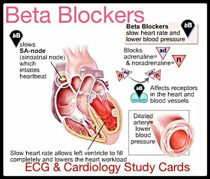
The most common form of treatment is a medication given orally called βeta blockers. This helps reduce the intensity of the heart's work, help to prevent the heart from beating too fast and can control arrhythmias easing the strain on the cardiac structures. Another form of treatment is to reduce the workload on the heart to decrease the chance of sudden cardiac arrest, fainting or death. This entails regulating the dog's exercise and preventing sudden increases in activity and intense exertion.
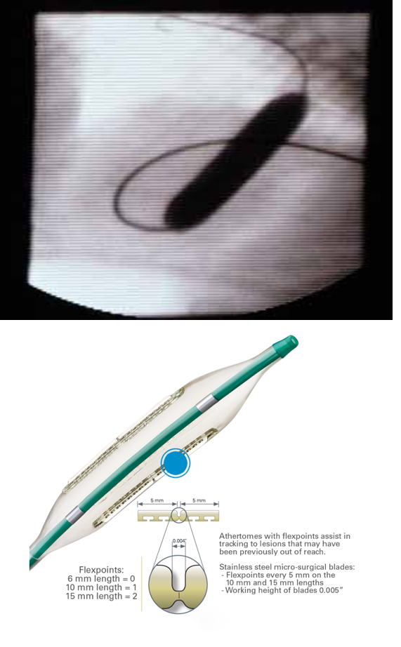
Aside from drugs, there are several surgical procedures that reduce the obstruction of subaortic stenosis but with varied successes. A minimally invasive technique of balloon catheterization/ valvuloplasty involves inserting a catheter down the jugular vein into the heart and inflated in the vessel or chamber where the obstruction is occurring. It opens up the lumen of the vessel providing immediate relief from the strain on the heart. Recently a new technique has become available utilizing a special "cutting" balloon. The advantages if this balloon compared to the original balloon used is that the cutting balloon has multiple arthrotomes attached to it. These microsurgical blades are able to make incisions in the coronary segment during inflation increasing the vessel diameter in a more controlled manner. It also requires a lower inflation pressure than the conventional balloon inflation which could reduce the possibility of vessel wall injury.


References:


Comments